How Gut Bacteria Changes the Immune System

By Dr. Narveen Jandu  

The human immune system changes with age. Immune responses start to become less robust as people get older, which makes them more vulnerable to certain infections and diseases.

However, immune system aging looks different from person to person. Research has shown that changes to the composition and diversity of the microorganisms in the gut may explain these differences in immune system aging.

The gut microbiome — the population of microorganisms that lives in the gastrointestinal tract — helps the body maintain a stable internal environment when it is faced with external changes. This is known as homeostasis. The gut microbiome supports homeostasis in different ways, such as through helping to keep the immune system alert, and digesting dietary fibre into short-chain fatty acids to strengthen the intestinal wall.

The gut microbiome also helps us to regulate our inflammatory reactions. Inflammation helps the body fight microorganisms that cause disease, and helps repair damaged tissues. However, as the composition of our gut microbiome changes with age, a low level of inflammation can become constant throughout the body. This is called inflammaging.

When inflammaging develops in the gut, it leads to a decrease in immune responses, which puts people at a higher risk for infection and disease.

Let’s take a closer look at the gut microbiome and how it changes with age.

Diversity of Bacteria Decreases with Age

Our gastrointestinal tract can be compared to a densely populated city inhabited by a variety of different bacteria, fungi, archaea and viruses collectively called the gut microbiota. In fact, compared to other parts of the body, the gut microbiome has the largest number of bacteria. In a healthy gut microbiome, there are four dominant families (or phyla) of microorganisms, including Firmicutes, Bacteroidetes, Proteobacteria and Actinobacteria.

Firmicutes and Bacteroidetes make up around 80 to 90 per cent of the gut microbiota in the digestive tract. Firmicutes help with the production of short-chain fatty acids to support intestinal health and the secretion of mucus to improve intestinal wall defence. Bacteroidetes metabolize complex carbohydrates into vitamins and nutrients, and help promote glycogen storage to improve glucose metabolism.

The gut microbiome and immune system work closely together. The microorganisms in the gut send out signals that are detected by immune sensors. This allows the immune system to regulate the beneficial bacteria in the gut, helping maintain immune homeostasis. Through this interaction, the adaptive immune system also receives stimuli from harmful substances called antigens, which trigger an immune reaction.

However, as people age, the composition and balance of microorganisms in the gut changes. This gives rise to microbial dysbiosis, which means there is a reduction in the number of beneficial bacteria in the gut, alongside a higher number and pro-inflammatory organisms and bacteria that can cause disease. In addition to this, research has also shown that the general diversity of bacteria in our gut also decreases with age.

Over time, the shortage of beneficial bacteria such as Firmicutes in older adults starts to compromise the integrity of their intestinal barrier, causing it to become leaky. This is because the Firmicutes family plays a very important role in keeping the intestinal wall healthy and strong by producing a short-chain fatty acid called butyrate. Short-chain fatty acids such as butyrate help provide nutrients to strengthen the intestinal wall, inform immune responses and lower inflammation.

When intact, the intestinal barrier works to prevent harmful bacteria from passing through the intestinal wall, entering the circulatory system and reaching important organs. However, when there are not enough gut bacteria to produce the short-chain fatty acids that are needed for the intestinal wall to function, bacteria are able to enter the bloodstream. This contributes to the formation of intestinal inflammaging, which refers to a low level of inflammation that becomes steady throughout the body with age.

Inflammaging creates an environment that is prone to inflammation, which is caused and maintained by several factors. These can include microorganism imbalances in the intestines (microbial dysbiosis), psychological stress, physical inactivity, poor nutrition and chronic infections.

When the body is exposed to these factors on a regular basis, cellular senescence occurs. Cellular senescence is a state in which cell growth is permanently arrested, which means that cells are no longer able to self-renew. Eventually, this leads to a decrease in immune responses, which are important to prevent foreign substances and pathogens from entering the body.

How to Maintain a Healthy Balance of Bacteria

There is a common saying that claims “you are what you eat.” Indeed, nutrition and diet play an important role in regulating the number of different microorganisms that live in the gut. This means that diet may also play a key role in the immune function of older adults.

The Mediterranean diet, known for its lower intake of refined carbohydrates, saturated fats, dairy products and red meat, has been shown to have a positive effect on the balance of microorganisms in the gut and the strength of the intestinal barrier. The Mediterranean diet has also been linked to a lower risk of Type 2 diabetes in older adults, allowing these individuals to live a longer and healthier life.

The use of probiotics and prebiotics can also help fight age-related inflammation. Probiotics, such as Lactobacilli and Bifidobacteria, are live microorganisms that can be consumed to support overall health. More specifically, probiotics help improve the function of the intestinal barrier and regulate immune responses by modifying the composition of the gut microbiome. However, there is still some debate around whether the acidic conditions in the stomach allow probiotics to survive long enough to be able to move into the intestine.

It is clear that the immune system has an intricate relationship with the gut microbiome. A healthy and well-balanced gut microbiome will strengthen the intestinal barrier, which helps to reduce inflammation throughout the body and support the immune system.

To achieve this, it is important to maintain a healthy and well-balanced lifestyle as we grow older. This can include lower intake of dairy products and red meats, and harnessing the benefits of probiotics and prebiotics.

Narveen Jandu, PhD, is a faculty member with the School of Public Health Sciences at the University of Waterloo in Canada. As a biomedical researcher, Dr. Jandu’s research has focused on studying the cellular mechanisms and pathophysiological consequences of infectious diseases.

This article was co-authored by Flore Van Leemput. a student in health sciences at the University of Waterloo.

This article originally appeared in The Conversation and is republished with permission.

How Inflammation Can Lead to Chronic Pain

By Drs. Prakash Nagarkatti and Mitzi Nagarkatti, University of South Carolina

When your body fights off an infection, you develop a fever. If you have arthritis, your joints will hurt. If a bee stings your hand, your hand will swell up and become stiff. These are all manifestations of inflammation occurring in the body.

We are two immunologists who study how the immune system reacts during infections, vaccination and autoimmune diseases where the body starts attacking itself.

While inflammation is commonly associated with the pain of an injury or the many diseases it can cause, it is an important part of the normal immune response. The problems arise when this normally helpful function overreacts or overstays its welcome.

Generally speaking, the term inflammation refers to all activities of the immune system that occur where the body is trying to fight off potential or real infections, clear toxic molecules or recover from physical injury. There are five classic physical signs of acute inflammation: heat, pain, redness, swelling and loss of function. Low-grade inflammation might not even produce noticeable symptoms, but the underlying cellular process is the same.

Take a bee sting, for example. The immune system is like a military unit with a wide range of tools in its arsenal. After sensing the toxins, bacteria and physical damage from the sting, the immune system deploys various types of immune cells to the site of the sting. These include T cells, B cells, macrophages and neutrophils, among other cells.

The B cells produce antibodies. Those antibodies can kill any bacteria in the wound and neutralize toxins from the sting. Macrophages and neutrophils engulf bacteria and destroy them. T cells don’t produce antibodies, but kill any virus-infected cell to prevent viral spread.

Collateral Damage

Additionally, these immune cells produce hundreds of types of molecules called cytokines – otherwise known as mediators – that help fight threats and repair harm to the body. But just like in a military attack, inflammation comes with collateral damage.

The mediators that help kill bacteria also kill some healthy cells. Other similar mediating molecules cause blood vessels to leak, leading to accumulation of fluid and influx of more immune cells.

This collateral damage is the reason you develop swelling, redness and pain around a bee sting or after getting a flu shot. Once the immune system clears an infection or foreign invader – whether the toxin in a bee sting or a chemical from the environment – different parts of the inflammatory response take over and help repair the damaged tissue.

After a few days, your body will neutralize the poison from the sting, eliminate any bacteria that got inside and heal any tissue that was harmed.

Inflammation is a double-edged sword. It is critical for fighting infections and repairing damaged tissue, but when inflammation occurs for the wrong reasons or becomes chronic, the damage it causes can be harmful.

Allergies, for example, develop when the immune system mistakenly recognizes innocuous substances – like peanuts or pollen – as dangerous. The harm can be minor, like itchy skin, or dangerous if someone’s throat closes up.

Chronic inflammation damages tissues over time and can lead to many noninfectious clinical disorders, including cardiovascular diseases, neurodegenerative disorders, obesity, diabetes and some types of cancers.

The immune system can sometimes mistake one’s own organs and tissues for invaders, leading to inflammation throughout the body or in specific areas. This self-targeted inflammation is what causes the symptoms of autoimmune diseases such as lupus and arthritis.

Another cause of chronic inflammation that researchers like us are currently studying is defects in the mechanisms that curtail inflammation after the body clears an infection.

While inflammation mostly plays out at a cellular level in the body, it is far from a simple mechanism that happens in isolation. Stress, diet and nutrition, as well as genetic and environmental factors, have all been shown to regulate inflammation in some way.

There is still a lot to be learned about what leads to harmful forms of inflammation, but a healthy diet and avoiding stress can go a long way toward helping maintain the delicate balance between a strong immune response and harmful chronic inflammation.

Prakash Nagarkatti, PhD, and Mitzi Nagarkatti, PhD, are Professors of Pathology, Microbiology and Immunology at the University of South Carolina. They receive funding from the National Science Foundation and the National Institutes of Health.

This article originally appeared in The Conversation and is republished with permission.

Anti-Inflammatory Drugs May Contribute to Chronic Pain

By Pat Anson, PNN Editor

Anti-inflammatory drugs that are widely used to treat short-term acute pain disrupt the body’s natural healing process and increase the chances of developing chronic pain, according to a provocative new study by an international team of researchers.

If true, it means that ibuprofen, naproxen, diclofenac and other non-steroidal anti-inflammatory drugs (NSAIDs) used by millions of people every day for the temporary relief of acute pain may contribute to long-term pain that is even harder to treat.    

“For many decades it’s been standard medical practice to treat pain with anti-inflammatory drugs. But we found that this short-term fix could lead to longer-term problems,” said co-author Jeffrey Mogil, PhD, Professor of Pain Studies and the Canada Research Chair in the Genetics of Pain at McGill University in Montreal.

In a series of studies, Mogil and his colleagues first analyzed genes and immune cells in the blood of 98 patients with acute lower back pain (LBP), noting which patients became free of pain and which ones developed chronic pain after three months.  

In patients who became pain free, there was an early inflammatory response to acute pain that activated neutrophils -- a type of white blood cell that helps the body fight infection.

In patients with chronic pain, there was no inflammatory immune response. This suggests that neutrophils play an active role in resolving pain and protecting patients from transitioning to chronic pain.

“Neutrophils dominate the early stages of inflammation and set the stage for repair of tissue damage. Inflammation occurs for a reason, and it looks like it’s dangerous to interfere with it,” said Mogil.

The study findings, published in Science Translational Medicine, were replicated in a cohort of patients with temporomandibular disorder (TMD), a painful inflammation of the jaw.

Researchers also tested their theory on laboratory animals, giving mice with acute pain the anti-inflammatory steroid dexamethasone or the NSAID diclofenac. While the drugs were initially effective, researchers found that blocking neutrophils in mice ultimately prolonged their pain up to ten times the normal duration. Three other analgesics without anti-inflammatory properties (gabapentin, morphine and lidocaine) produced short-term pain relief without affecting the overall duration of pain in mice.

These findings were also supported by a separate analysis of health records for 500,000 people in the United Kingdom with acute LBP. Those that took NSAIDs were nearly twice as likely to still have pain 2 to 6 years later than those who did not take NSAIDs. Patients who took acetaminophen (paracetamol) or antidepressants – neither of which are anti-inflammatory --  were not at higher risk of transitioning to chronic LBP.

“Our findings suggest it may be time to reconsider the way we treat acute pain. Luckily pain can be killed in other ways that don’t involve interfering with inflammation,” said co-author Massimo Allegri, MD, Head of Pain Service at Policlinico of Monza Hospital in Italy and Ensemble Hospitalier de la Cote in Switzerland.

Researchers say their findings should be followed up with larger clinical trials directly comparing the long-term effects of anti-inflammatory drugs to other pain relievers that don’t disrupt inflammation.

“Together, our results suggest that active immune processes confer adaptation at the acute pain stage, and impairment of such inflammatory responses in subjects with acute LBP (or TMD) increases the risk of developing chronic pain. These adaptive inflammatory responses are intrinsically transcriptionally driven, probably modified by both genetics and environmental factors, and can be inhibited by steroids and NSAIDs,” researchers said.

“Our conclusions may have a substantial impact on medical treatment of the most common presenting complaints to health care professionals. Specifically, our data suggest that the long-term effects of anti-inflammatory drugs should be further investigated in the treatment of acute LBP and likely other pain conditions.”

NSAIDs are widely used to treat everything from fever and headache to low back pain and arthritis. They are in so many different pain relieving products, including over-the-counter cold and flu medications, that many consumers may not be aware how often they use NSAIDs. At high doses, studies have found that NSAIDs increase the risk of a heart attack or stroke.

The current draft revision of the CDC opioid guideline recommends that NSAIDs should be used for low back pain, painful musculoskeletal injuries, dental pain, postoperative pain, kidney stones and acute pain caused by episodic migraine.

Acetaminophen also has its risks. Long-term use has been associated with liver, kidney, heart and blood pressure problems. Acetaminophen overdoses are involved in about 500 deaths and over 50,000 emergency room visits in the U.S. annually.

Natural Herbs for Intractable Pain Syndrome

By Dr. Forest Tennant, PNN Columnist 

Intractable Pain Syndrome (IPS) is defined as constant pain with cardiovascular, metabolic and hormonal complications. IPS is caused by neuroinflammation inside the brain and spinal cord (central nervous system or CNS) that comes from excess electromagnetic energy generated by a painful disease or injury.

Excess electromagnetic energy activates an immune cell in the CNS called “microglia” to produce inflammation that then destroys tissue in the CNS. Unfortunately, tissue destroyed by inflammation impairs or damages the normal CNS mechanisms that shut off or cause pain to cease. A person may, therefore, develop constant (24/7) pain that overstresses the cardiovascular, metabolic and hormonal systems.

Tissue destruction in the CNS is well documented by brain scans. This relatively recent understanding of how neuroinflammation destroys CNS tissues and causes constant pain is arguably the most important discovery for pain treatment in the 21st Century. Why? We now have some ideas on how to treat IPS that can possibly cure or at least permanently reduce pain rather than just provide temporary, symptomatic relief. 

Treating CNS Inflammation 

When someone develops IPS, it is human nature to seek immediate pain relief and ignore its basic cause. If you have constant (24/7) pain, however, one must accept the fact that you have inflammation in the CNS that must be suppressed. Otherwise, you can reasonably assume that the pain will get worse.  

While research has documented that CNS inflammation may spread, it is unknown whether it ever “burns out.” As of yet, there is no blood or x-ray test to know if “burn out” may occur. This means that every person with IPS must take one or more anti-inflammatory agents to stop further tissue destruction and the worsening of pain. 

A problem when suppressing inflammation in the CNS is that only a few of the anti-inflammatory agents which are commercially available cross the blood brain barrier and enter the spinal fluid in sufficient amounts to be effective. This includes the non-specific anti-inflammatory drugs (NSAIDs) and corticosteroids.  

Benefits of Natural Herbal Products

Interestingly, natural products such as botanicals, herbs, enzymes and hormones tend to cross the blood brain barrier and provide anti-inflammatory activity. A well-known common example is aspirin (acetylsalicylic acid), which is derived from willow tree bark.

This has caused a great deal of interest in the use of natural products for suppression of CNS inflammation. Several research studies in both laboratory tests and animals have found that some natural agents do indeed suppress CNS inflammation.

To date, research has identified five herbs that suppress CNS inflammation. There are likely other natural products that suppress CNS inflammation, but this list is a good start:

  1. Ginseng

  2. Curcumin

  3. Resveratrol

  4. Ginger

  5. Fisetin

Currently, there are very few controlled blind studies in humans to demonstrate the effectiveness of these herbal products. Personally, I have often seen considerable effectiveness of natural products in reducing the pain of IPS. Other anecdotal reports from patients and doctors are also starting to accumulate.

Precise dosages are unknown, but the manufacturer of each herbal product will have some starting instructions on the label. Herbal agents appear quite safe and have few reported side effects. Herbs can be taken with corticosteroids, opioids, naltrexone, electrical stimulators, neuropathic agents, and essentially all medication used for treatment of IPS.

At this time, we believe there is enough research and clinical experience to recommend both herbal, non-prescription as well as prescription anti-inflammatory agents to assist treatment of IPS. The time has come to treat IPS with a broader-based approach rather than just the use of symptomatic pain relievers.

Based on our current knowledge, IPS will likely get worse unless a person’s treatment program includes agents that suppress CNS inflammation.

Forest Tennant, MD, DrPH, MPH is retired from clinical practice but continues his research on the treatment of arachnoiditis and intractable pain at the Arachnoiditis Research and Education Project. The Tennant Foundation gives financial support to Pain News Network and sponsors PNN’s Patient Resources section.  

Exercise Reduces Pain by Increasing Beneficial Bacteria

By Pat Anson, PNN Editor

Regular exercise can benefit people in many different ways, helping us lose weight, reduce the risk of heart disease, and boosting overall health.  

But researchers at the University of Nottingham have found that exercise has an unexpected benefit for people with arthritis. Regular exercise increases levels of beneficial bacteria in their digestive tracts, which reduces pain and inflammation by increasing levels of endocannabinoids – cannabis-like substances naturally produced by the body.

The study, published in the journal Gut Microbes, is believed to be the first to find a potential link between endocannabinoids, exercise and gut microbes.

"Our study clearly shows that exercise increases the body's own cannabis-type substances. Which can have a positive impact on many conditions,” says lead author Amrita Vijay, a Research Fellow at Nottingham’s School of Medicine. "As interest in cannabidiol oil and other supplements increases, it is important to know that simple lifestyle interventions like exercise can modulate endocannabinoids."

Vijay and her colleagues enrolled 78 people in their study. Half of the participants did 15 minutes of muscle strengthening exercises every day for six weeks, and the rest did nothing. Blood and fecal samples were collected from both groups.

At the end of the study, participants who exercised not only had lower pain levels, they also had significantly more Bifidobacteria and Coprococcus 3 -- bacteria that produce anti-inflammatory substances and lower levels of cytokines, which regulate inflammation.

These gut bacteria were particularly adept at raising levels of short chain fatty acids (SCFAs), which increase levels of endocannabinoids. About a third of the anti-inflammatory effects of the gut microbes was due to their ability to raise endocannabinoid levels.

Importantly, the exercise group also had lower levels of Collinsella – a bacteria known to increase inflammation that is strongly associated with processed food and diets low in vegetables.    

“In this study we show that circulating levels of ECs (endocannabinoids) are consistently associated with higher levels of SCFAs, with higher microbiome diversity and with lower levels of the pro-inflammatory genus Collinsella. We also show statistically that the anti-inflammatory effects of SCFAs are up to one third mediated by the EC system,” researchers concluded.

Previous studies have also found an association between gut bacteria and painful conditions. A 2019 study at McGill University found that women with fibromyalgia had 19 different species of bacteria that were present in either greater or lesser quantities than a healthy control group.

Bacteria associated with irritable bowel syndrome, chronic fatigue syndrome and interstitial cystitis were also found to be abundant in the fibromyalgia patients, but not in the control group.    

Having a healthy diet can also affect pain levels for migraine, neuropathy and other types of chronic pain. A recent study funded by the National Institutes of Health found that migraine sufferers who ate more fatty fish and reduced their consumption of polyunsaturated vegetable oils — frequently found in processed foods — had fewer headaches.

Inflammation Cascades Cause Intractable Pain Syndrome

By Forest Tennant, PNN Columnist

The greatest research breakthrough in recent years that will provide much hope for persons with intractable pain syndrome (IPS) has been the discovery of the “inflammatory cascades” inside the central nervous system (CNS). Now that this is known, there are specific measures to take to reduce or halt this process.

Most persons with IPS have two inflammatory cascades, one in the brain and the other in the lower spinal canal. They are called cascades because one area of inflammation can ignite another in a continuous chain reaction and spread.

Excess electric currents enter the brain from a bodily site of damage or diseases. In response to tissue injury, inflammation initially begins around receptors and spreads to other parts of the brain, just like the inflammation that causes cellulitis on the skin.

Inflammation in the lower spinal canal usually starts in a protruding intervertebral disc or due to a puncture, infection or injury to the dura-arachnoid covering of the spinal canal.

Once inflammation starts in either the brain or inside the spinal canal, it may not ever cease or burn-out. It may even silently continue to spread and damage CNS tissue with progressive pain and disability.

How to Control Inflammation

While pain relief is always the first thing on the mind of an afflicted person, measures to control and suppress the inflammatory cascade are essential. Normal anti-inflammatory agents don’t usually enter the brain or spinal canal in adequate amounts to be very helpful. You must use multiple agents that cross the blood brain barrier.

Every person with IPS must be on a daily program that include three measures to control the cascades of CNS inflammation or your condition will likely deteriorate. This can be done through diet, vitamins and supplements that help prevent inflammation; medications that reduce inflammation; and movements that help keep spinal fluid flowing.

Review the three measures listed here with your family and medical practitioners. Select the nutrients, medications and exercises to develop a program that best fits you.

You must continue daily cascade control for as long as you have pain. Opioids and other analgesics, by themselves, do not control the inflammatory cascades. Without a cascade control program, you can expect that your pain relievers will diminish in effectiveness and may totally stop working.

Forest Tennant is retired from clinical practice but continues his research on intractable pain and arachnoiditis. This column is adapted from newsletters recently issued by the IPS Research and Education Project of the Tennant Foundation. Readers interested in subscribing to the newsletter can sign up by clicking here.

The Tennant Foundation gives financial support to Pain News Network and sponsors PNN’s Patient Resources section.   

Is Your Spinal Pain Inflammatory or Neuropathic?

By Forest Tennant, PNN Columnist

Every person with Adhesive Arachnoiditis (AA) or other spinal canal disorder needs to determine if their pain is primarily inflammatory, neuropathic or both. Why? The treatments are different.

AA is fundamentally an inflammatory disease that involves two different intraspinal canal tissues: the cauda equina nerve roots and the arachnoid-dural covering of the spinal canal. The inflammation causes damage to the nerve roots, so electricity either can’t pass or it doesn’t pass in a smooth, natural flow.

Nerve damage that blocks or alters electricity conduction is called “neuropathic” pain. AA usually has both inflammatory and neuropathic pain, but the inflammation may resolve and leave behind damaged nerve roots and neuropathic pain.

The inflammatory and neuropathic pain of AA may also develop into Intractable Pain Syndrome, which is constant, incurable pain with cardiovascular, endocrine (hormonal) and autoimmune complications.

Persons with AA usually need to treat both kinds of pain – inflammatory and neuropathic --   but one type may be predominant. A blood test for inflammatory markers is helpful, but not totally diagnostic.

If your pain improves with a trial of ketorolac (1 or 2 injections) or a corticosteroid (Medrol Dose Pak or dexamethasone), you have active inflammation that must be treated. We also recommend botanical anti-inflammatory agents, such as curcumin/turmeric, Andrographis and serrapeptase.

Prescription medications for neuropathic pain include gabapentin (Neurontin), diazepam, carisoprodol, topiramate, Lyrica and Cymbalta.

Every person with AA of the cervical and/or lumbar spines should experiment with topical medications, such as the Salonpas patch, lidocaine gel or patch, Voltaren gel and diclofenac (prescription needed).

Topical medication that is applied and massaged into the skin may dissolve through the tissues to the inflamed or damaged area. On average, you can expect 10 to 25% additional pain relief, plus the potential to permanently reduce your pain. Sometimes topical  medication will relieve painful areas that other drugs taken orally or by injection cannot reach.

Forest Tennant is retired from clinical practice but continues his research on the treatment of intractable pain and arachnoiditis. This column is adapted from bulletins recently issued by the Arachnoiditis Research and Education Project. Readers interested in subscribing to the bulletins should send an email to tennantfoundation92@gmail.com.

The Tennant Foundation gives financial support to Pain News Network and sponsors PNN’s Patient Resources section.  

Injections of Tiny Particles Reduce Osteoarthritis Knee Pain

By Pat Anson, PNN Editor

A minimally invasive procedure significantly reduces pain and inflammation caused by knee osteoarthritis, according to preliminary research being presented this week at the annual meeting of the Society of Interventional Radiology.

Geniculate artery embolization (GAE) is a relatively new procedure in which thousands of microscopic particles are injected into arthritic knees. The particles reduce inflammation by disrupting the abnormal flow of blood caused by osteoarthritis (OA), a joint disorder that causes thinning of cartilage and progressive joint damage. As the cartilage breaks down, it releases enzymes that cause inflammation and pain.

GAE takes about one to two hours, and many patients with knee OA report significant improvement in pain and physical function that can last up to a year.

"Prior to treatment, patients' knee pain had taken over their whole life," said lead researcher Siddharth Padia, MD, a professor of radiology at UCLA Health. "But after treatment, patients who initially could walk only three or four blocks were walking three miles. Some were able to do away with walking aids, such as canes, while others reported being in a better mood now that they were living without pain."

For their Phase 2 study, Padia and his colleagues enrolled 40 patients with knee OA who were not candidates for total knee replacement, and who failed to benefit from pain relievers, joint injections and physical therapy.

Catheters were inserted into arteries leading to the knees through pinhole incisions in the patients’ hips. The microscopic particles — called Embozene microspheres — were then slowly injected through the catheter into the knees. Each patient was evaluated for pain and adverse events at one week; one, three and six months; and one year after the treatment.

Researchers say patients saw benefits as soon as three days after the procedure. Average pain levels decreased from 8 out of 10 before GAE to 3 out of 10 within the first week. Most patients reported more than 50% reduction in their pain levels at the one-year follow up.

Adverse events, such as skin ulceration and small bone infarction – the death of bone tissue due to reduced blood supply -- were reported by 9 patients, but resolved without treatment.

Embozene microspheres are made by Boston Scientific and are currently used in the treatment of vascular tumors, uterine fibroids and arterial malformations. They must be carefully injected into affected tissue to prevent them from circulating in the blood and reaching healthy tissue and organs.

“This prospective trial demonstrates that GAE is highly effective and durable in reducing symptoms due to moderate to severe knee OA that is refractory to other conservative therapy, and has an acceptably low toxicity profile,” researchers concluded.

The UCLA researchers plan to conduct a larger, randomized trial to determine which patients may benefit most from GAE and the impact it has on slowing the progression of arthritis.

Results from other studies on the use of GAE are also being presented at the meeting of the Society of Interventional Radiology. One review found that GAE can be effective for patients who don't respond well to conservative treatments for knee OA, but cautioned that “definitive conclusions can't be made on the true efficacy of GAE until studies are done with longer follow up and larger patient numbers.”

How to Reduce Brain and Spinal Cord Inflammation

By Forest Tennant, PNN Columnist

Intractable pain syndrome (IPS) is constant pain with cardiovascular and endocrine dysfunction. IPS occurs when the initial cause of pain creates inflammation in the brain and spinal cord. This is called neuroinflammation.

Inflammation in the brain and spinal cord is what causes the worsening of IPS symptoms. Inflammation does its dirty work by burning out or damaging neurotransmitter systems such as dopamine, endorphin, cannabinoid, serotonin, and gaba aminobutyric acid (GABA). Common symptoms of neuroinflammation:

  • Constant pain

  • Fatigue

  • Amotivation (Lack of motivation or purpose)

  • Attention deficit

  • Memory impairment

  • Elevated blood pressure & pulse

  • Social withdrawal

  • Dietary change

  • Weight gain

  • Sugar craving

  • Depression

Every person with IPS must attempt to control and reduce their brain and spinal cord inflammation. To reduce neuroinflammation, we recommend regular consumption of one or more of these non-prescription, natural herbal medicinal agents:

  • Tumeric/Curcumin

  • Ashwagandha

  • Boswellia

  • Palmitoyethanolamine (PEA)

  • Traumeel

  • Cannabidiol (CBD)

  • Andrographis

You can take any of these on different days or several together, as long as you use at least one daily.

If the disorder that started your pain and IPS ends in “itis” -- arthritis, arachnoiditis, pancreatitis, cystitis, colitis or myositis -- you will also need a periodic (e.g., 1-2 times a week) low dose of a corticosteroid such as hydrocortisone, methylprednisolone, prednisone or dexamethasone.

Don’t rely on pain relievers alone. You must have an inflammation reduction component as part of your IPS treatment program.

Forest Tennant is retired from clinical practice but continues his research on intractable pain and arachnoiditis. This column is adapted from newsletters recently issued by the IPS Research and Education Project of the Tennant Foundation. Readers interested in subscribing to the newsletter can sign up by clicking here.

The Tennant Foundation has given financial support to Pain News Network and sponsors PNN’s Patient Resources section.  

New Drug Could Improve Effectiveness of Stem Cell Therapy

By Pat Anson, PNN Editor

Scientists have developed an experimental drug that can lure stem cells to damaged tissues and help them heal -- a discovery being touted as a major advancement in the field of regenerative medicine.

The findings, recently published in the Proceedings of the National Academy of Sciences (PNAS), could improve the effectiveness of stem cell therapy in treating spinal cord injuries, stroke, amyotrophic lateral sclerosis (ALS), Parkinson’s disease and other neurodegenerative disorders. It could also expand the use of stem cells to treat conditions such as heart disease and arthritis. 

“The ability to instruct a stem cell where to go in the body or to a particular region of a given organ is the Holy Grail for regenerative medicine,” said lead author Evan Snyder, MD, director of the Center for Stem Cells & Regenerative Medicine at Sanford Burnham Prebys Medical Discovery Institute in La Jolla, CA. “Now, for the first time ever, we can direct a stem cell to a desired location and focus its therapeutic impact.”

Over a decade ago, Snyder and his colleagues discovered that stem cells are drawn to inflammation -- a biological “fire alarm” that signals tissue damage has occurred. However, using inflammation as a therapeutic lure for stem cells wasn’t advisable because they could further inflame diseased or damaged organs, joints and other tissue.

To get around that problem, scientists modified CXCL12 -- an inflammatory molecule that Snyder’s team discovered could guide stem cells to sites in need of repair— to create a drug called SDV1a. The new drug works by enhancing stem cell binding, while minimizing inflammatory signals.

“Since inflammation can be dangerous, we modified CXCL12 by stripping away the risky bit and maximizing the good bit,” Snyder explained. “Now we have a drug that draws stem cells to a region of pathology, but without creating or worsening unwanted inflammation.”

To demonstrate its effectiveness, Snyder’s team injected SDV1a and human neural stem cells into the brains of mice with a neurodegenerative disease called Sandhoff disease. The experiment showed that the drug helped stem cells migrate and perform healing functions, which included extending lifespan, delaying symptom onset, and preserving motor function for much longer than mice that didn’t receive the drug. Importantly, the stem cells also did not worsen the inflammation.

Researchers are now testing SDV1a’s ability to improve stem cell therapy in a mouse model of ALS, also known as Lou Gehrig’s disease, which is caused by a progressive loss of motor neurons in the brain. Previous studies conducted by Snyder’s team found that broadening the spread of neural stem cells helps more motor neurons survive — so they are hopeful that SDV1a will improve the effectiveness of neuroprotective stem cells and help slow the onset and progression of ALS. 

“We are optimistic that this drug’s mechanism of action may potentially benefit a variety of neurodegenerative disorders, as well as non-neurological conditions such as heart disease, arthritis and even brain cancer,” says Snyder. “Interestingly, because CXCL12 and its receptor are implicated in the cytokine storm that characterizes severe COVID-19, some of our insights into how to selectively inhibit inflammation without suppressing other normal processes may be useful in that arena as well.”

Snyder’s research is supported by the National Institutes of Health, U.S. Department of Defense, National Tay-Sachs & Allied Disease Foundation, Children’s Neurobiological Solutions Foundation, and the California Institute for Regenerative Medicine (CIRM).

“Thanks to decades of investment in stem cell science, we are making tremendous progress in our understanding of how these cells work and how they can be harnessed to help reverse injury or disease,” says Maria Millan, MD, president and CEO of CIRM. “This drug could help speed the development of stem cell treatments for spinal cord injury, Alzheimer’s, heart disease and many other conditions for which no effective treatment exists.”

Great Progress Being Made in Treating Arachnoiditis

By Dr. Forest Tennant, PNN Columnist

About 5 years ago, most medical practitioners had either never heard of Adhesive Arachnoiditis (AA) or thought it was a spider bite. Today, almost all practitioners in the modern world have heard of AA. Many now understand it and some even treat it. A few are trying some innovative new approaches.

AA is a chronic inflammation that starts inside the spinal canal that can lead to severe suffering, neurologic impairments and a shortened lifespan. Once inflammation starts, it apparently never, or rarely, goes totally away.  

Treatment and prevention in recent years have greatly reduced the occurrence of some serious neurologic impairments and autoimmune complications of AA. The most obvious decrease in new cases reviewed by the Tennant Foundation are those of upper and lower extremity paraparesis (partial paralysis) and total paralysis, which are rapidly disappearing.

Urinary and bladder impairments that require catheterization are also hardly seen. And the autoimmune manifestations of arthritis, thyroid deficiency and carpal tunnel are disappearing.

Why the improvement? Awareness, thanks to patients, social media and advocates who have educated the medical profession about AA. Fewer epidurals, early treatment and emergency measures have all helped. The development of protocols for prevention, emergency intervention and on-going treatment have been essential.

Major Remaining Problems

Persons with AA are still having difficulty, in some communities, finding medical practitioners who are comfortable and willing to treat AA. The major complication is the development of constant pain and the intractable pain syndrome.

The key to preventing AA and stopping its progression is early treatment. Our research has clearly shown that AA is almost always preceded by one of 3 intraspinal canal inflammatory conditions:

  1. Protruding, degenerated intravertebral discs.

  2. Cauda equina inflammation.

  3. Arachnoid inflammation (i.e. plain arachnoiditis) due to collagen disorders or needle injury.

Some intraspinal canal inflammatory disorders always precede AA. These disorders should be aggressively treated to prevent AA.

Select Corticosteroids Essential for AA

We believe all persons with typical AA symptoms and documentation of the disease on an MRI must take one of two corticosteroids (CS): methylprednisolone or dexamethasone for the spinal canal inflammation and pain of AA.

Currently there is no other medication agent that consistently and predictably suppresses intraspinal canal inflammation and reduces pain. Do not expect to halt progression or have much recovery if you do not consistently take a CS.

Dexamethasone and methylprednisolone are the preferred CS’s because they cross the blood brain barrier, enter spinal fluid and act on glial cells. Prednisone and hydrocortisone are not as consistently effective as dexamethasone and methylprednisolone, which should be taken in low doses.

  1. Maintenance-low dose of dexamethasone (.5 to .75mg) or methylprednisolone (Medrol) 2 to 4 mg on 2 to 5 days a week. Skip days between dosages. An alternative is a weekly or bi-monthly injection of methylprednisolone or dexamethasone. Injections are usually the answer to corticoid sensitivity or gastric upset.

  2. For flares, a 6-Day Medrol Dose Pak or an injection of methylprednisolone or dexamethasone, preferably mixed with a standard dose of injectable ketorolac.

The fear of corticosteroids comes from daily use of high doses, not from low, intermittent dosages. Some persons with severe asthma and rheumatoid arthritis must take a corticosteroid for years and don’t experience serious side effects.

Forest Tennant, MD, MPH, DrPH, is retired from clinical practice but continues his groundbreaking research on the treatment of intractable pain and arachnoiditis. This column is adapted from bulletins recently issued by the Arachnoiditis Research and Education Project of the Tennant Foundation. Readers interested in subscribing to Dr. Tennant’s bulletins should send an email to tennantfoundation92@gmail.com.

Dr. Tennant and the Tennant Foundation have given financial support to Pain News Network and are currently sponsoring PNN’s Patient Resources section.  


New Imaging Better Identifies Sources of Chronic Pain

By Pat Anson, PNN Editor

A new approach to diagnostic imaging that combines the use of positron emission tomography (PET) and magnetic resonance imaging (MRI) could help identify the causes of chronic pain better than current methods, according to a new study at Stanford University School of Medicine.

PET scans that identify increased glucose metabolism in the body – known as 18F-FDG PET -- are currently used by oncologists to detect where tumors are located. Stanford researchers say the same approach could also be used to more precisely locate inflammation in injured nerves and muscles.

"In the past few decades, we have confirmed that anatomic-based imaging approaches, such as conventional MRI, are unhelpful in identifying chronic pain generators," said Sandip Biswal, MD, an associate professor of radiology at Stanford University School of Medicine.

"We know that 18F-FDG PET has the ability to accurately evaluate increased glucose metabolism that arises from to acute or chronic pain generators. As such, in our study we examined PET/MRI as a potential solution to determine the exact molecular underpinnings of one's pain."

In the study, 65 chronic pain patients underwent 18F-FDG PET and MRI scans from head to toe. The PET/MR images were then evaluated by two radiologists to determine if more glucose uptake occurred in painful areas or in other parts of the body. Increased glucose metabolism was detected in 58 of the 65 patients.

One such patient was an adult male who lived with decades of chronic pain in his neck as a result of an injury experienced at birth. PET/MR scans — such as the image on the right — identified muscles in his right neck near a spinal nerve where there was elevated glucose uptake.

This finding encouraged a surgeon to explore the area. The surgeon found a collection of small arteries wrapped around the nerve. When the arteries were ablated (removed) by the surgeon, it relieved pressure on the nerve and the patient reported tremendous relief.

Cipriano, et al., Stanford University

Cipriano, et al., Stanford University

As a result of PET/MR imaging, 40 patients in the study had their pain management plans modified, including some who had procedures that their doctor had not anticipated, such as surgery to relieve foot pain and the placement of blood patches to treat cerebrospinal fluid leaks.

"The results of this study show that better outcomes are possible for those suffering from chronic pain," said Biswal. "This clinical molecular imaging approach is addressing a tremendous unmet clinical need, and I am hopeful that this work will lay the groundwork for the birth of a new subspecialty in nuclear medicine and radiology.”

Biswal recently presented his findings at the annual meeting of the Society of Nuclear Medicine and Molecular Imaging. They’ve also been published in the Journal of Nuclear Medicine.

Tennant Foundation Launches Intractable Pain Research and Education Project

By Pat Anson, PNN Editor

One of the pioneers of pain management in the United States is hoping to draw more attention to intractable pain and how it differs from chronic pain. While chronic pain lasts for 90 days or more, intractable pain can persist for years, decades or even a lifetime. Many doctors fail to distinguish between the two, which leads to gaps in diagnosis and treatment.

“The pain field has been woefully negligent about this. They talk about symptomatic treatment of what to do to relieve chronic pain, but in order to deal with intractable pain syndrome you’ve got to treat the underlying cause,” says Dr. Forest Tennant, whose foundation has just released a new report called “The Intractable Pain Syndrome: A Call for Recognition and Prevention.”

“We’ve worked on it for quite some time with the goal of having every patient, doctor, nurse and family understand it. And to try and bring things out in language that is understandable by everybody.”

“This was a labor of love for all of us, born from a sincere desire to bring recognition, treatment and prevention to this devastating syndrome,” says co-author Ingrid Hollis, whose son was treated by Tennant for Arachnoiditis and Ehlers-Danlos Syndrome. “We have learned so much through the years, and I believe there is much that can be done to prevent intractable pain syndrome from developing and progressing when it occurs.”

“One of the reasons we’re doing this is to call for early recognition and vigorous treatment,” Tennant told PNN. “Someone who has this for two, five or twenty years, you can’t expect much reversal. But those people who have these conditions for six months or a year, they have a great ability to reverse a great deal of this. The earlier the treatment, the better the outcome.”

Left untreated, Tennant says intractable pain syndrome (IPS) can lead to chronic inflammation in the central nervous system that is difficult to reverse.  The inflammation not only causes constant pain, it leads to loss of brain tissue and creates dysfunction in the neurologic, cardiovascular, hormone and immune systems.  

“We’ve known for centuries that a painful injury will create what amounts to electricity. And too much electricity causes inflammation and inflammation causes tissue destruction,” says Tennant. “Intractable pain syndrome does change their basic physiology by virtue of altering physically the neurotransmitter systems that are in the brain and spinal cord. That is really the difference. And this is very objective. It’s measurable. It’s visible. It’s clear when its present.”

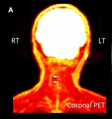
Tennant says these “pockets” of inflammation can be seen on MRI’s and brain scans. Someone suffering from simple chronic pain won’t have them, but people with intractable pain will.

There are five basic conditions that can cause IPS:

  1. Arachnoiditis, a chronic inflammation of spinal nerves

  2. Genetic connective tissue/collagen disorders such as Ehlers-Danlos Syndrome (EDS)

  3. Reflex Sympathetic Dystrophy (RSD), also known as Complex Regional Pain Syndrome (CRPS)

  4. Brain injuries caused by strokes or head trauma

  5. Serious end-stage osteoarthritis of the spine, hips, knees or feet.

Other possible causes of IPS are porphyria, sickle cell disease, Lyme disease, interstitial cystitis, and some rare genetic disorders.   

IPS Can Be Reversed

Long recognized as incurable, Tennant says intractable pain can be reversed if diagnosed and treated early. He has pioneered some new treatments for IPS, including hormone supplements, physical therapy and better nutrition.

“Our project hopes to educate people about what they can do to reverse a lot of this,” Tennant told PNN. “Step number one is you’ve got to have vigorous treatment of the underlying cause of pain. The arachnoiditis has to be treated. The arthritis has to be treated. The RSD has to be treated. In other words, vigorous treatment of the underlying condition.

“Step number two, there are specific things they can do to slow down the inflammation inside the central nervous system. We know the body makes hormones inside the brain and spinal cord that can reduce the inflammation inside the central nervous system and regrow some of the tissue that’s been lost. People say you can’t do that, but oh yes, you can. There are studies that show this and we see this clinically.”

“I strongly believe that Dr. Tennant's concept of how intractable pain develops and progresses is absolutely correct.  His work in identifying the causes, developing diagnostic criteria, and figuring out a treatment protocol provides what doctors need to know to help people with intractable pain,” says co-author Kristen Ogden, whose husband Louis was treated by Tennant for a complex autoimmune condition.

“Louis' treatment with Dr. Tennant allowed him to improve so much in so many ways. Very effective pain control, greatly improved function and the best quality of life he ever had as an adult.  I am sure that very few people have any idea at all how much a seriously ill pain patient can improve and even regain lost function and capabilities if they have the right medical regimen that meets their needs.”

Tennant retired from clinical practice in 2018, the year after his home and office were raided by the DEA as part of an investigation into his opioid prescribing. No charges were ever filed against him. While Tennant no longer sees patients, he is continuing his research into the treatment of IPS – which notably downplays the use of opioids.

“The answer to opioid use is treating the syndrome rather than just giving symptomatic relief. Opioids are symptomatic drugs,” explains Tennant. “What we’re saying is, identify this and try to treat the condition. Don’t just throw a bunch of symptomatic antidepressants, opioids and other interventions at it. Start trying to look at it physiologically and pathologically, and treat it like we do other syndromes.”    

To learn more about the Tennant Foundation’s Intractable Pain Syndrome Research and Education Project, click here. 

Dr. Tennant and the Tennant Foundation have given financial support to Pain News Network and are currently sponsoring PNN’s Patient Resources section.  

How to Avoid the Holiday Blues

By Pat Anson, PNN Editor

For many of us, the holiday season wouldn’t be complete without Christmas cookies, jelly donuts, plum pudding, chocolate babka, or even the much-maligned fruit cake.

But if you're prone to depression or have an inflammatory condition, you might want to avoid those sweet treats. Or at least enjoy them in moderation.

New research by clinical psychologists at the University of Kansas suggests that dietary sugars found in many holiday foods can trigger metabolic, inflammatory and neurobiological processes that can lead to insomnia, digestive problems and depression – which all enhance physical pain.

"A large subset of people with depression have high levels of systemic inflammation,” said lead author Stephen Ilardi, PhD, an associate professor of clinical psychology at KU. "When we think about inflammatory disease we think about things like diabetes and rheumatoid arthritis - diseases with a high level of systemic inflammation. We don't normally think about depression being in that category, but it turns out that it really is.

“We also know that inflammatory hormones can directly push the brain into a state of severe depression. So, an inflamed brain is typically a depressed brain. And added sugars have a pro-inflammatory effect on the body and brain."

Depression Causes Sugar Cravings

Dwindling daylight in winter can worsen depression and prompt people to consume more sweets, which provide a temporary emotional lift.

"One common characteristic of winter-onset depression is craving sugar," Ilardi said. "So, we've got up to 30% of the population suffering from at least some symptoms of winter-onset depression, causing them to crave carbs - and now they're constantly confronted with holiday sweets.

"When we consume sweets, they act like a drug. They have an immediate mood-elevating effect, but in high doses they can also have a paradoxical, pernicious longer-term consequence of making mood worse, reducing well-being, elevating inflammation and causing weight gain."

The KU research team analyzed a wide range of studies on the physiological and psychological effects of sugar, including the Women's Health Initiative study, the NIH-AARP Diet and Health Study, and studies of Australian and Chinese soda-drinkers. Their research is published in the journal of Medical Hypotheses.

Ilardi says consuming high amounts of sugar could be as physically and psychologically harmful as drinking too much liquor.

"We have pretty good evidence that one alcoholic drink a day is safe, and it might have beneficial effect for some people," Ilardi said. "Alcohol is basically pure calories, pure energy, non-nutritive and super toxic at high doses. Sugars are very similar."

The average American gets about 14% of their calories from added sugars – the equivalent of 18 teaspoons of sugar each day. Most people know a high-sugar diet can lead to diabetes, obesity and cardiovascular problems.

Another way to look at sugar is to think of it as fuel for bacteria.  

"Our bodies host over 10 trillion microbes and many of them know how to hack into the brain," Ilardi said. "The symbiotic microbial species, the beneficial microbes, basically hack the brain to enhance our well-being. They want us to thrive so they can thrive.

“But there are also some opportunistic species that can be thought of as more purely parasitic - they don't have our best interest in mind at all. Many of those parasitic microbes thrive on added sugars, and they can produce chemicals that push the brain in a state of anxiety and stress and depression. They're also highly inflammatory."

Ilardi recommends eating a minimally processed diet rich in fruits, vegetables, fish and whole grains, while avoiding red meats, refined grains, fructose and other unhealthy foods. As for sugar, the KU researcher urges moderation -- not just during the holidays, but year-round.

Can Melatonin Put Your Chronic Pain to Sleep?

By A. Rahman Ford, PNN Columnist

Melatonin is popularly known as the sleep hormone. Less known is its potential to alleviate chronic pain and inflammation.

Melatonin is a natural hormone produced by the pineal gland in the brain. During the day the pineal gland is inactive, but at night it begins to produce melatonin and helps us sleep.

As a supplement, melatonin is widely promoted for its efficacy as a sleep aid. However, its role in reducing inflammation – a major contributor to chronic pain – may be much more important. Many chronic pain conditions are a result of underlying inflammation.

In a recent Nature article, melatonin was called a “master regulator” of inflammation. Several studies have shown that melatonin can regulate activation of the immune system, reducing chronic and acute inflammation.

Research shows that melatonin supplements can modulate inflammation by acting as powerful antioxidants and free radical scavengers. Uncontrolled free radicals in the body can lead to oxidative stress, which can cause inflammation and culminate in diseases that cause chronic pain.

There is a large body of evidence that melatonin is a potent antioxidant, even more potent than vitamins C and E.  It’s been successfully used to treat fibromyalgia and irritable bowel syndrome, diseases associated with high levels of oxidative stress.

Melatonin is also a strong antimicrobial, and emerging research shows that some chronic inflammatory conditions may be caused by infections. One study found melatonin effective in treating certain drug-resistant bacteria such as Staphylococcus aureus, Pseudomonas aeruginosa and Acinetobacter baumannii. Similar results were found when testing ten different pathogens, including Escheria coli and Salmonella typhinurium, with melatonin.

Melatonin and Chronic Pain

A therapeutic role for melatonin in the treatment of painful autoimmune conditions has been theorized.  A 2013 study noted that melatonin plays a role in the pathogenesis of conditions such as multiple sclerosis and systemic lupus. In animal models of these diseases, melatonin supplements were found to have protective effects. A 2019 review concurred, concluding that melatonin can serve as a new therapeutic target in treating autoimmune diseases.

A review of the scientific literature on chronic pain syndromes found evidence of melatonin’s efficacy as an analgesic in several conditions including fibromyalgia, irritable bowel syndrome and chronic back pain. Studies also showed melatonin’s effectiveness in treating cluster headaches ad tension headaches.

A small clinical trial of 63 females with fibromyalgia found that melatonin, alone or in combination with the antidepressant amitriptyline, significantly reduced pain when compared to amitriptyline use alone. The authors concluded that the melatonin treatment had a direct effect on the regulation of pain.

There has been some evidence that melatonin supplements can help reduce lower back pain. In a 2015 study, researchers found a significant reduction in pain intensity during movement and at rest in patients with back pain.

Melatonin has also been successful in treating migraines. In an open-labeled clinical trial of 34 patients suffering from migraine, 30 mg of melatonin given 30 minutes before bedtime was found to reduce headache intensity as well as frequency and duration, with significant clinical improvement after one month.

Although the scientific evidence is only slowly emerging, melatonin is a widely-available, inexpensive and safe supplement that may aid you in your fight against chronic pain.

A. Rahman Ford, PhD, is a lawyer and research professional. He is a graduate of Rutgers University and the Howard University School of Law, where he served as Editor-in-Chief of the Howard Law Journal.

Rahman lives with chronic inflammation in his digestive tract and is unable to eat solid food.

The information in this column is for informational purposes only and represent the author’s opinions alone. It does not inherently express or reflect the views, opinions and/or positions of Pain News Network.