Lack of Awareness Is Harming People with Scoliosis  

By Drs. Sanja Schreiber and Emily Somers

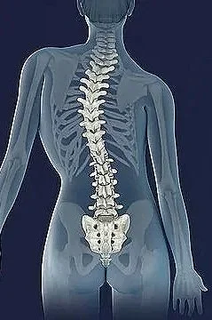
Cael was a typical 15-year-old — until the discovery of an already advanced abnormal curvature of his spine.

“I felt like the Hunchback of Notre Dame,” Cael told CBC News, recalling the emotionally draining and gruesome two-year wait for spinal surgery during which his curve progressed to a whopping 108 degrees.

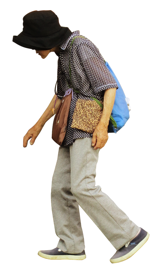
Scoliosis is an abnormal twisting and curving of the spine that can develop at any age, but mostly occurs during rapid growth spurts in children, and as part of spine aging in adults over the age of 60.

Of all types of scoliosis in children, adolescent idiopathic scoliosis is the most prevalent, accounting for as many as nine in 10 cases and impacting up to one in 20 adolescents globally. On the other end of the age spectrum, a staggering two-thirds of older adults are also affected.

In clinical care, research and education related to scoliosis, disparities persist worldwide. Despite its widespread prevalence, scoliosis often goes undiagnosed, or has delayed diagnosis as in Cael’s case. It also receives limited attention in clinical and public health education, leading to significant gaps in health care.

This general lack of awareness has serious implications for thousands of people like Cael.

Gaps in Scoliosis Care

In the United States, fewer than half of states legislate school-based scoliosis screening in children. Even worse, Canada discontinued screening back in 1979 because it was not considered cost-effective.

Pediatricians’ screening practices vary, and some cases of scoliosis in children are only discovered when an unrelated chest X-ray reveals a curved spine. With about 30 per cent of cases being hereditary, parents may not recognize the signs early on.

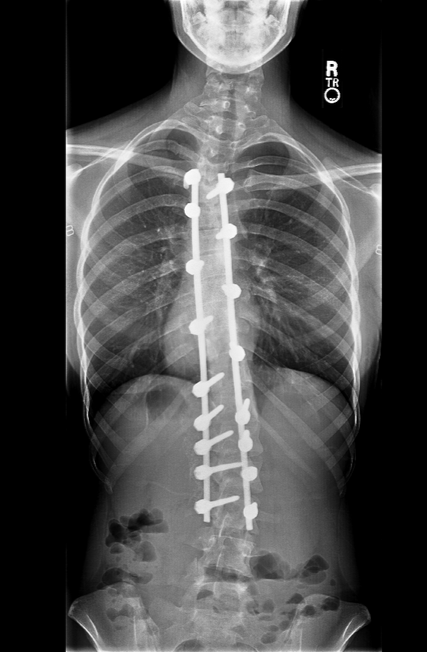
The recommended care in North America involves bracing for mild to moderate curves (25° to 45°) and surgery for curves exceeding 45°. Shockingly, 32 per cent of Canadian children, like Cael, face delayed referrals, discovering significant curves when they finally see specialists.

Despite documented success in managing scoliosis through early screening, exercise rehabilitation and brace treatment, global health-care education often neglects this condition.

The general lack of global awareness leaves physicians, nurses and other practitioners unaware of effective treatments and referral processes, contributing to the misunderstanding and under-treatment of patients. Consequently, when children with scoliosis eventually reach specialists for care, they may encounter challenges navigating the health-care system as they transition into adulthood.

Sex Disparities

It is unclear why adolescent idiopathic scoliosis affects mainly girls. The more severe the curve, the more likely the patient is female.

Due to their specific biology, females also face a five-fold higher risk of progressive deformities and are 10 times more likely, compared to males, to require surgery.

Despite generally uncomplicated pregnancies and deliveries, women with scoliosis often face difficulties receiving pain control during labour, with higher epidural failure rates.

Moreover, they often suffer pregnancy-related back pain, and their spine curvature may worsen after pregnancy.

Healthcare Barriers

Healthcare access in the U.S. is influenced by a range of factors including race, income and health insurance coverage.

Patients with better insurance plans tend to seek pediatric orthopedic care at a younger age. Those with public insurance tend to have worse spine curvatures by the time they reach a scoliosis specialist; this is particularly striking among Black patients with public insurance, who are 67 per cent less likely to be diagnosed at a stage early enough for effective brace treatment compared to Black patients with private insurance.

While Canada’s health-care system covers spinal fusion for severe scoliosis, the lack of a national insurance program in the U.S. leads to varying out-of-pocket expenses for patients.

Those without insurance often cannot afford surgery at all.

But even with Canada’s universal coverage, patients typically wait an entire year for surgery due to a shortage of providers. Because of regional variability in resources such as access to spinal surgeons, funding and specialized facilities, some kids, like Cael, wait even longer, experiencing physical, emotional and psychological burdens, while their curves get progressively worse.

Delayed surgery in Canada cost the health-care system $44.6 million due to more complex surgeries, extended hospital stays, readmission and re-operation rates.

Lack of Research

Ongoing gender disparities in the healthcare workforce and lack of research funding for this female-predominant condition continue to hamper effective action.

Fewer than five per cent of spinal surgeons identify as women. Glass-ceiling effects surround women surgeons in this male-dominated culture, perpetuating gendered training environments, being held to higher standards and earning lower wages. The dearth of senior women role models and mentors is a further barrier for career advancement and retention.

Furthermore, research funding for diseases, such as scoliosis, that mainly affect females has historically lagged far behind funding for male-predominant diseases. Improving workforce diversity is an important facet of addressing health disparities and shaping research agendas.

Inequities abound in scoliosis care and research. The impact of lack of awareness and delayed care extends beyond physical challenges. The patient and their family suffer emotionally, incurring significant financial burden while fearing the future.

The message is clear, we must do better for this underserved population.

Sanja Schreiber, PhD, is an Adjunct Professor of Physical Therapy at the University of Alberta and the Owner/Director of Curvy Spine, a specialized clinic for treating structural spinal disorders and training physiotherapists in scoliosis rehabilitation.

Emily Somers, PhD, is an epidemiologist specializing in immune-mediated and musculoskeletal diseases. She also directs the Interdisciplinary Research & Team Science Program at the Michigan Institute for Clinical and Health Research.

This article originally appeared in The Conversation and is republished with permission.
Stem Cell Discovery Could Revolutionize Spine and Cancer Care

By Pat Anson, PNN Editor

The discovery of a new type of stem cell could revolutionize the treatment of spine disorders and slow the progression of some cancers, according to a groundbreaking study published in Nature.

Researchers from Hospital for Special Surgery (HSS) and Weill Cornell Medicine say the vertebral stem cells they found in human spines appear to play a key role in spinal health and in the metastasis of cancerous tumors as they spread through the body.

“There are two big takeaway discoveries that were made here. One is that we have discovered a stem cell that forms the spine and maintains the spine throughout life. This cell makes all the other cells that mineralize the spine,” said lead investigator Matthew Greenblatt, MD, associate professor of pathology and laboratory medicine at Weill Cornell Medicine.

“The second discovery here is that we found that this stem cell drives tumors. Breast cancer is what we focused on here, but likely also prostate cancer.”

Cancer experts have long believed that tumors metastasize to other parts of the body simply through blood flow. But Greenblat and his colleagues found that vertebral stem cells essentially attract cancer cells to the spine. That could explain why some cancers are first detected in the spine after they have metastasized from the breast, prostate and lung.

“Because we found that molecular ‘come here’ signal that's made by this spine stem cell, that gives us the ability to block that signal therapeutically. And that's something we're working on to try to prevent or treat established spine metastases,” Greenblatt told PNN.

Boosting Bone Health

Researchers say their discovery could also lead to breakthroughs in spinal health, by giving physicians a way to speed up recovery from spinal injuries and slow the progression of degenerative conditions such as osteoporosis, a disease that makes bones thinner, less dense and more likely to fracture.

For example, someone with degenerative disc disease could have their vertebral stem cells harvested, reproduced in a laboratory, and then reinjected to stimulate the growth of new bone. In animal tests, human vertebral stem cells helped laboratory mice form new bones in their spines.

“We can show that they formed basically little vertebral bones when those patient cells are put into mice, which really tells us that we found the right cell. And we can work with the cell transplant and retain stability to make new bone,” said co-author Sravisht Iyer, MD, a spine surgeon at HSS.

“I think kind of figuring out how to recruit the cells or how to how to encourage them to form more bone is going to be an important area or avenue of investigation for us, as a way to help people and protect people against what is a very morbid condition for them.”

Iyer says early treatment with vertebral stem cells could help someone with osteoporosis or a spine fracture, but wouldn’t necessarily benefit patients suffering from more advanced cases of bone loss.

“By the time people are presenting to us with spine pain, they usually have some element of compressive pathology or a degree of degeneration, which will likely require some intervention, whether that's surgery or epidural injection,” Iyer said.

“Where this work I think can really help push us forward is once you get those at-risk patients, they probably will need a surgery because a lot of degeneration is asymptomatic, and by the time they get to you they probably need something, but maybe you can prevent the second, third or fourth operation or intervention.”

More research and human studies are needed before the stem cells can be used in clinical settings to improve bone health and slow the metastasis of cancer cells. But researchers are excited by what they’ve learn so far.    

“We predict this discovery will lead to the targeting of these cells to disrupt the function and ultimately reduce the spread of cancer to the spine," said Greenblatt.

My Arachnoiditis Family

By Elaine Ballard, Guest Columnist

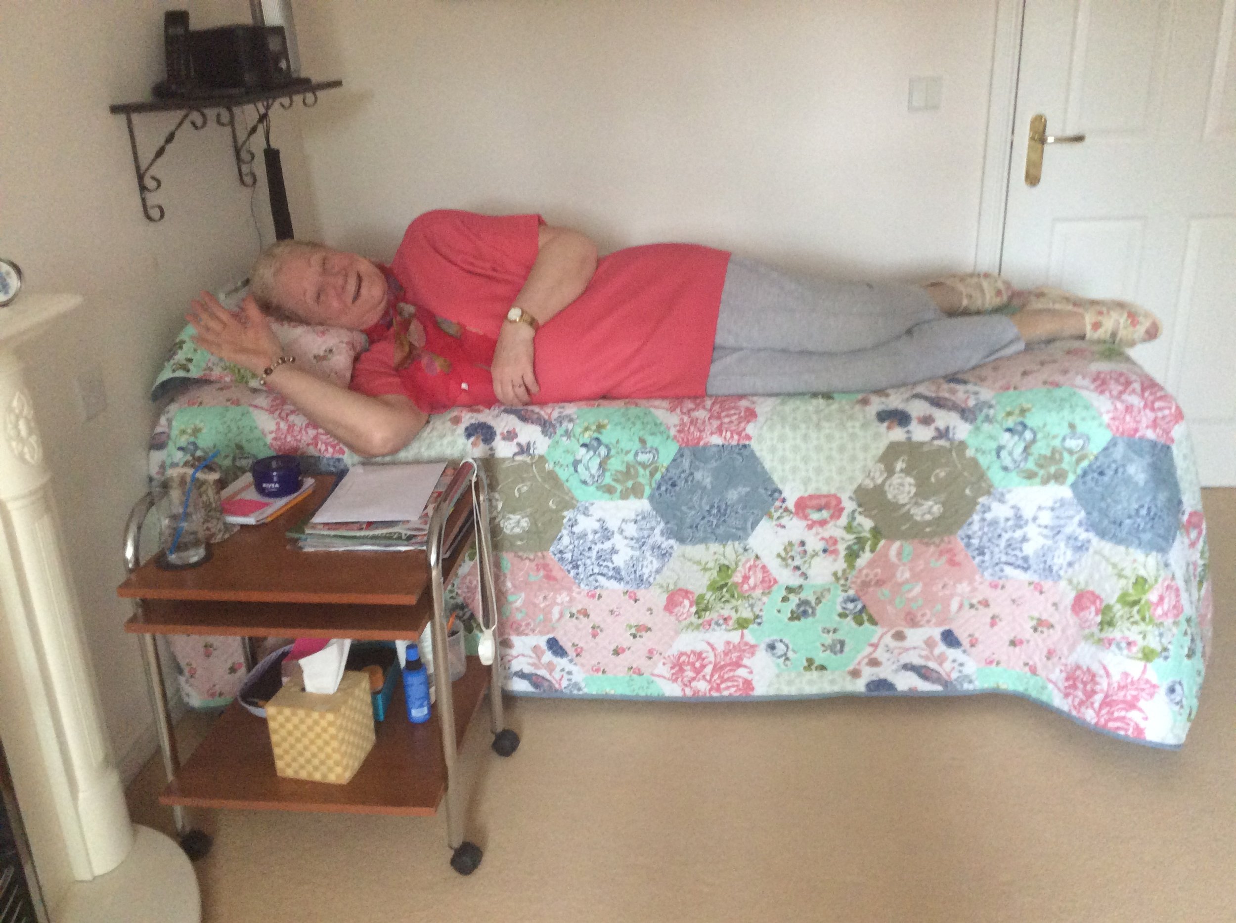
I live in the rural county of Somerset in England, UK. At the age of 22, I had a sporting accident which eventually left me 80 percent disabled and unable to lead a normal life. 

The accident caused several crushed discs in my spine and a great deal of nerve damage. Over the years multi-level disc degeneration set in, as well as osteoarthritis. I am unable to use a wheelchair, as bulging discs prevent me from sitting without severe pain. I am now 73.

ELAINE BALLARD

Since 1994, I have been confined to lying on a bed in my living room and only leave home to keep hospital appointments. I travel by stretcher ambulance.

Just over two years ago I had an MRI scan which showed I had Adhesive Arachnoiditis (AA) and my life changed drastically yet again. 

Arachnoiditis is listed as a rare neurological condition, but in fact many thousands of people all over the world have been diagnosed with it. There are also thousands of other people who have the same symptoms, but as yet, no diagnosis.

It is difficult for patients to get diagnosed as doctors are not trained to recognize this disease and often fail to even recognize the symptoms.

Arachnoiditis results from severe inflammation of the arachnoid membrane that surrounds the nerves of the spinal cord. It may cause stinging and burning pain, as well as muscle cramps, spasms, and uncontrollable twitching. The most common symptom is severe to unbearable neurological pain, especially to the nerves connecting to the lower back, legs and feet. This can lead to tingling, numbness, weakness and severe pain in the legs and feet.

Other symptoms include sensations that feel like insects crawling on the skin or water trickling down the legs. It can also affect the bladder, bowel and sexual function. Unfortunately for some, it may also result in paralysis.

As this disease progresses, the symptoms can become more severe or even permanent. Most people with Arachnoiditis are eventually unable to work and suffer significant disability because they are in constant pain. Pain is the most dominant factor and it is both chronic and acute. As the disease progresses, it can be relentless and unbearable and sadly suicide becomes an option.

Inflammation of the arachnoid membrane can lead to the formation of scar tissue, which may cause the spinal nerves to clump together and eventually adhere to the lining wall of the dura, the middle layer of the spine. The disease can then progress to Adhesive Arachnoiditis.

What Causes Arachnoiditis

There are a few different causes of Arachnoiditis. In the 1970's a dye used in myelograms was injected during spinal procedures directly into the area surrounding the spinal cord and nerves. The dye was too toxic for these delicate parts of the spine and was blamed for causing Arachnoiditis. This dye continues to be used in some parts of the world.

Bacterial infections and viruses in the spine can also lead to Arachnoiditis. So can complications from spinal surgery and invasive spinal procedures such as epidural steroid injections.

There is no cure for Arachnoiditis and there is little effective pain relief. This is a disease or condition for life. Opioids are offered by doctors, but are not specific to reducing neurological pain of this nature.

It is very sad and cruel that opioids are being clamped down in America and that Arachnoiditis patients are being classed together with people who seek drugs for recreational purposes. We are not drug seekers but desperate victims crying out for something that will stop this relentless and overwhelming neurological pain.

The Facebook support group Arachnoiditis Together We Fight has been an important part of my education in understanding this disease. I am thankful to say it has become more of a family, where members can come in and gradually feel at home while we bring education, support and encouragement. This family atmosphere and great support has saved many lives, as people first arrive feeling suicidal and lost in a medical world that will not help them.

That is why I wrote this poem to show people how important support groups can be and to bring more attention to this rare but life changing disease.

"The Family"

By Elaine Ballard

Lonely, fearfully I knock at the door
Arac greets me, a smile, so kind
I want to die, eyes keep to the floor
"Welcome" she says, but what will I find?

"Welcome" repeated again and again
"Good to have you!" Are you kidding?  
"Family" really can it be true? 
Lost, lonely, rejected... what you too? 

I tell my story, they will never believe
"We understand, you're not alone"
Tears trickle down, I cannot believe
We are bound together by this dreaded disease

Files, inflammation, medication
Head's in a spin, where do I begin?  
Then a hand upon my shoulder
Guides me to those precious folders

Questions answered, hope is rising
Found some friends, pain subsiding
Flares still come but under control
No longer afraid nor out in the cold

We need each other, your pain is mine  
Strength in unity, love is the sign
Moving forward we are free
To Fight Together as one FAMILY


Elaine Ballard has written a book about Adhesive Arachnoiditis and how her Christian faith helped her through many difficult flares and times. It is called “The Furnace of Fire” and is available on Amazon. Click on the book's cover to see price and ordering information.

Pain News Network invites other readers to share their stories (and poems) with us. Send them to editor@painnewsnetwork.org.

The information in this column should not be considered as professional medical advice, diagnosis or treatment. It is for informational purposes only and represents the author’s opinions alone. It does not inherently express or reflect the views, opinions and/or positions of Pain News Network.

Spine and Hip Fractures Raise Risk of Chronic Body Pain

By Pat Anson, Editor

Breaking a bone in your spine or hip may be so traumatic that it doubles your chances of developing chronic widespread body pain such as fibromyalgia, according to a large new study by British researchers.

The study, published in the Archives of Osteoporosis, utilized an existing health database of over half a million adults to investigate associations between fractures of the spine, hip or upper and lower limbs, and the development later in life of chronic widespread body pain. Researchers at the University of Southampton also considered the possible effects of other factors, including diet, lifestyle, body build, and psychological health.

They found that men and women who had a spine fracture and women who had a hip fracture were more than twice as likely to experience long term widespread pain than those who did not have a fracture.

"The causes of chronic widespread pain are poorly characterized, and this study is the first to demonstrate an association with past fracture. If confirmed in further studies, these findings might help us to reduce the burden of chronic pain following such fractures," said lead researcher Nicholas Harvey, Professor of Rheumatology and Clinical Epidemiology at the University of Southampton.

"Chronic widespread pain is common, and leads to substantial health related problems and disability. Past studies have demonstrated an increased risk of chronic widespread pain following traumatic events, but none have directly linked to skeletal fractures."

Physical and emotional traumas have long been identified as risk factors for chronic widespread pain. For example, people involved in motor vehicle accidents are at greater risk of developing fibromyalgia, and rates of chronic widespread pain are known to increase after major disasters such as a hurricane or earthquake. Until now, there was no evidence that bone fractures could trigger such a response.

“Interestingly, the associations appeared strongest for fractures at the hip and spine, compared with fractures in the upper or lower limbs,” wrote Harvey. “High levels of morbidity and decreased survival following a hip and spine fractures is well documented, as are the potential changes in body shape, such as kyphosis, leading to pain and respiratory difficulties following vertebral fracture.”

Data for the research was collected from the UK Biobank study, which maintains records on almost everyone who utilizes the UK National Health Service.